Research Paper Spotlight: Detecting Proteins Associated with Alzheimer's
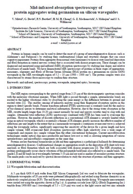
 The original research paper highlighted below, "Mid-infrared absorption spectroscopy of protein aggregates using germanium on silicon waveguides," can be found at the University of Southampton site: [external PDF link].
 In this paper, Dr. Vinita Mittal and her colleagues at the University of Southampton successfully used a quantum cascade laser to identify biological markers associated with Alzheimer's and Parkinson's diseases. Below are some highlights of their work.
In many neurodegenerative diseases, proteins in the body clump together, creating what are known as "amyloids" or "fibrils." These long-chain protein forms can accumulate in the brain, disrupting cellular function.
Although there is debate about whether amyloids and fibrils are the cause of Alzheimer's disease, symptoms often correlate with their presence. Therefore, detecting these types of protein structures can be a crucial step in early diagnosis and treatment of disease.
In their experiment, Dr. Mittal and her team began by developing fibrils in a Bovine Serum Albumin solution. To confirm the presence of fibrils, they initially used an Atomic Force Microscope to take images of:
- the ordinary "monomer" proteins,
- intermediate-phase "oligomers,"
- and finally, the ribbon-like fibrils.
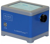
After confirming the three protein types through microscopy, the researchers then scanned each type using a LaserTune infrared tunable quantum cascade laser from Block Engineering.
They focused primarily on the Amide I (1600-1700 cm-1), Amide II (1480-1580 cm-1), and Amide III (1200-1300 cm-1) bands of the mid-infrared region. These wavelength ranges are particularly sensitive to the spectral signatures of proteins.
 In the resulting scans, they were able to detect measurable shifts in the absorption peaks and frequencies for each of the three protein forms. A clear distinction between the monomers, oligomers, and fibrils was apparent in the chemical signatures.
Although they cautioned that size and composition of the forms may account for some of the absorption differences, these results are nonetheless promising for early identification of protein markers in neurodegenerative diseases. The ability to differentiate healthy monomers from clumped fibrils may help in the detection of these diseases.
If you are a life science researcher interested in using tunable infrared lasers in your work, please contact us for information about how our QCLs can help.

