
Research Paper Spotlight: Studying Cancer Microenvironments using a QCL Confocal Microscope
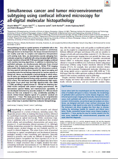 The original research paper highlighted below, "Simultaneous cancer and tumor microenvironment subtyping using confocal infrared microscopy for all-digital molecular histopathology," can be found at the Proceedings of the National Academy of Sciences (PNAS) site: [external web link].
In this paper, Dr. Rohit Bhargava and his colleagues at the University of Illinois at Urbana-Champaign describe the use of a laser-based confocal microscope to analyze cancer cells and the surrounding tissue microenvironment. Below are highlights of their work.
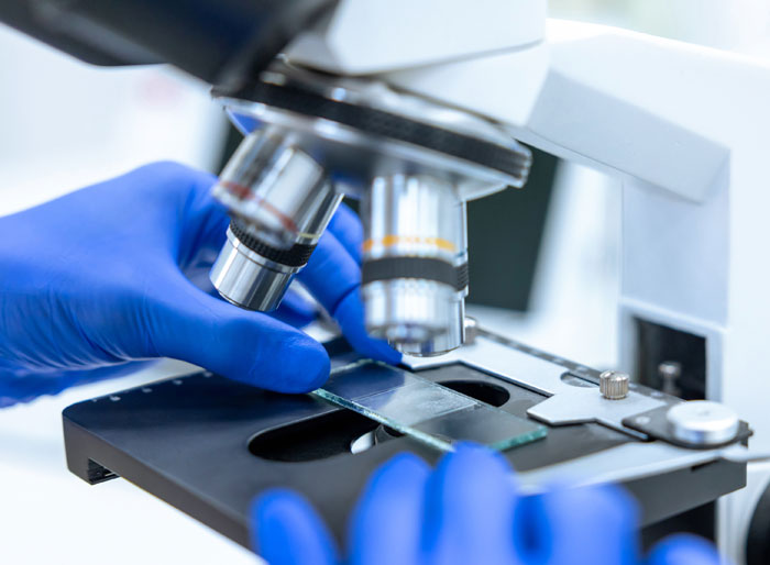
Traditionally, detection and analysis of cancer cells is accomplished by treating thin tissue samples with stains, and then examining color contrasts and other features under a conventional optical microscope.
 While this is an effective method for cancer detection, the approach doesn't allow for study of the surrounding tissue microenvironment. Neighboring "stromal" structures that may play a role in the development of tumors are generally excluded from this type of analysis.
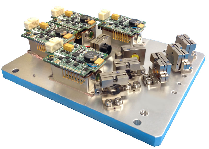
In order to allow for broader study of the cancer microenvironment, Dr. Bhargava and his associates built a new type of microscope powered by quantum cascade lasers from Block Engineering. The team's mid-infrared confocal microscope allows for detailed study of both epithelial and stromal tissues.
In their study, the researchers compared two technologies in the examination of breast tissue samples. The first approach used a commercial FTIR spectrometer connected to an imaging microscope. The second approach used the QCL confocal microscope that the team themselves developed.
The quality of the imaging scans from both instruments was excellent, and cancerous cells were clearly identified. However, the QCL confocal microscope produced results that were 50 times faster than the FTIR. This was largely due to the excellent signal-to-noise ratio and tunability of the infrared light. A set of scans that might require weeks in the FTIR setup could be completed in hours with the QCL confocal microscope.
 The laser-based microscope allowed researchers to bypass the tissue staining and manual observation approach used in traditional pathology. Data was collected digitally, and was available to be processed through algorithms and other computational frameworks. With the advent of AI-driven diagnostics, this type of data will be increasingly important.
Through the use of mid-infrared spectral imaging, the broader microenvironment of cancerous regions can be studied — including surrounding stromal tissue. The inclusion of tunable quantum cascade lasers in an imaging system allows for enormous speed advantages over FTIR instruments that have weaker globar light sources and poorer signal-to-noise ratios.
If you are a life science researcher interested in using QCLs in your work, please contact us for information about how our lasers can help.